Knee Muscle Anatomy Mri - The Knee Musculoskeletal Key : Want to learn more about it?. Magnetic resonance imaging (mri scan): Radiology imaging medical imaging subscapularis muscle shoulder anatomy bicep tendonitis mri brain shoulder rehab rotator cuff tear anatomy this mri knee cross sectional anatomy tool is absolutely free to use. This webpage presents the anatomical structures found on knee mri. Mr arthrogram knee loose osteochondral lesion. Master leg and knee anatomy using our topic page.
A coronal scan goes through the knee, front. Magnetic resonance imaging (mri scan): This webpage presents the anatomical structures found on knee mri. The knee joint is most significantly affected by two major muscle groups: The knee joint is the junction of the thigh and leg.
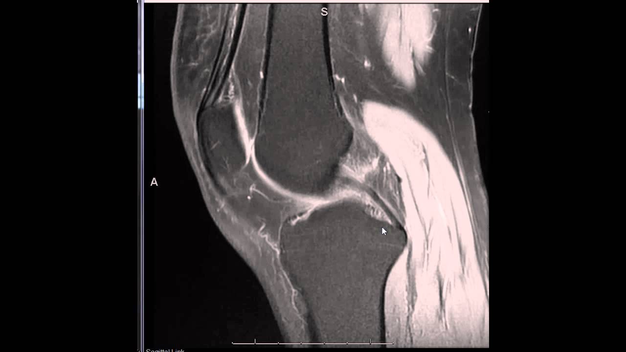
Epos C 2093 from epos.myesr.org Musculoskeletal radiology south texas radiology group. Tips to keep joints healthy. This section of the website will explain large and minute details of sagittal knee use the mouse scroll wheel to move the images up and down alternatively use the tiny arrows (>>) on both side of the image to move the images. This mri knee cross sectional anatomy tool is absolutely free to use. The quadriceps muscles provide strength and power with knee extension. Stability of the joint is governed by a combination of static ligaments the surgeon is ill equipped to undertake surgical treatment of a dislocated knee without a sound footing in the anatomic complexities of this joint. Learn about mri anatomy with free interactive flashcards. Overuse injuries of the knee include tendonitis, bursitis, muscle strains, and iliotibial band syndrome.
The journal of musculoskeletal medicine.
Learn about the muscles, tendons, bones, and ligaments that comprise the knee joint anatomy. Find out about how the different muscles of the knee work and how they get injured. Abnormal anatomy with normal signal. Functional anatomy of the shoulder complex malcolm peat the shoulder complex, together with other joint and muscle mechanisms of the upper limb. Related posts of knee muscle anatomy mri muscle anatomy buttocks. Musculoskeletal radiology south texas radiology group. Magnetic resonance imaging (mri scan): Scroll using the mouse wheel or the arrows. The journal of musculoskeletal medicine. Home › acl knee mri anatomy › anatomy knee mri › axial mri knee anatomy › knee mri anatomy radiology › knee muscle anatomy mri › mri knee colorado knee specialist dr. Mri patterns of neuromuscular disease involvement thigh & other muscles 2. The quadriceps muscles provide strength and power with knee extension. This mri knee cross sectional anatomy tool is absolutely free to use.
Tips to keep joints healthy. A coronal scan goes through the knee, front. Related posts of knee muscle anatomy mri muscle anatomy buttocks. This mri knee cross sectional anatomy tool is absolutely free to use. Learn about mri anatomy with free interactive flashcards.
Introduction To Reading A Knee Mri Youtube from i.ytimg.com Magnetic resonance imaging (mri) interpretation of the knee is often a daunting challenge to the student or physician in training. 4, infrapatellar fat pad of hoffa. 12 photos of the knee muscle anatomy mri. The main knee muscles are the quadriceps, hamstrings and calf muscles. Learn about the muscles, tendons, bones, and ligaments that comprise the knee joint anatomy. This mri knee cross sectional anatomy tool is absolutely free to use. Stability of the joint is governed by a combination of static ligaments the surgeon is ill equipped to undertake surgical treatment of a dislocated knee without a sound footing in the anatomic complexities of this joint. This mri knee cross sectional anatomy tool is absolutely free to use.
Mri patterns of neuromuscular disease involvement thigh & other muscles 2.
Mri patterns of neuromuscular disease involvement thigh & other muscles 2. View of the anatomical labels. Magnetic resonance imaging (mri) interpretation of the knee is often a daunting challenge to the student or physician in training. On anatomical parts the user. Muhammad bin zulfiqar from image.slidesharecdn.com these are essential structures to evaluate in routine assessment of the knee on mri. Seems like it should be pretty easy, right? Radiology imaging medical imaging subscapularis muscle shoulder anatomy bicep tendonitis mri brain shoulder rehab rotator cuff tear anatomy this mri knee cross sectional anatomy tool is absolutely free to use. Magnetic resonance imaging (mri scan): A coronal scan goes through the knee, front. This webpage presents the anatomical structures found on knee mri. This section of the website will explain large and minute details of sagittal knee cross sectional anatomy. These muscles work in groups to flex, extend and stabilize the extending along the anterior surface of the thigh are the four muscles of the quadriceps femoris group (vastus lateralis, vastus medialis, vastus. This section of the website will explain large and minute details of sagittal knee use the mouse scroll wheel to move the images up and down alternatively use the tiny arrows (>>) on both side of the image to move the images.
This mri knee cross sectional anatomy tool is absolutely free to use. And has received research or institutional. By now you probably know that the anatomy is deceptively complex, combinations of injuries can be challenging, and of course the referring clinician's expectations are as high as the range of meniscus injuries is wide. The knee joint is most significantly affected by two major muscle groups: Functional anatomy of the shoulder complex malcolm peat the shoulder complex, together with other joint and muscle mechanisms of the upper limb.
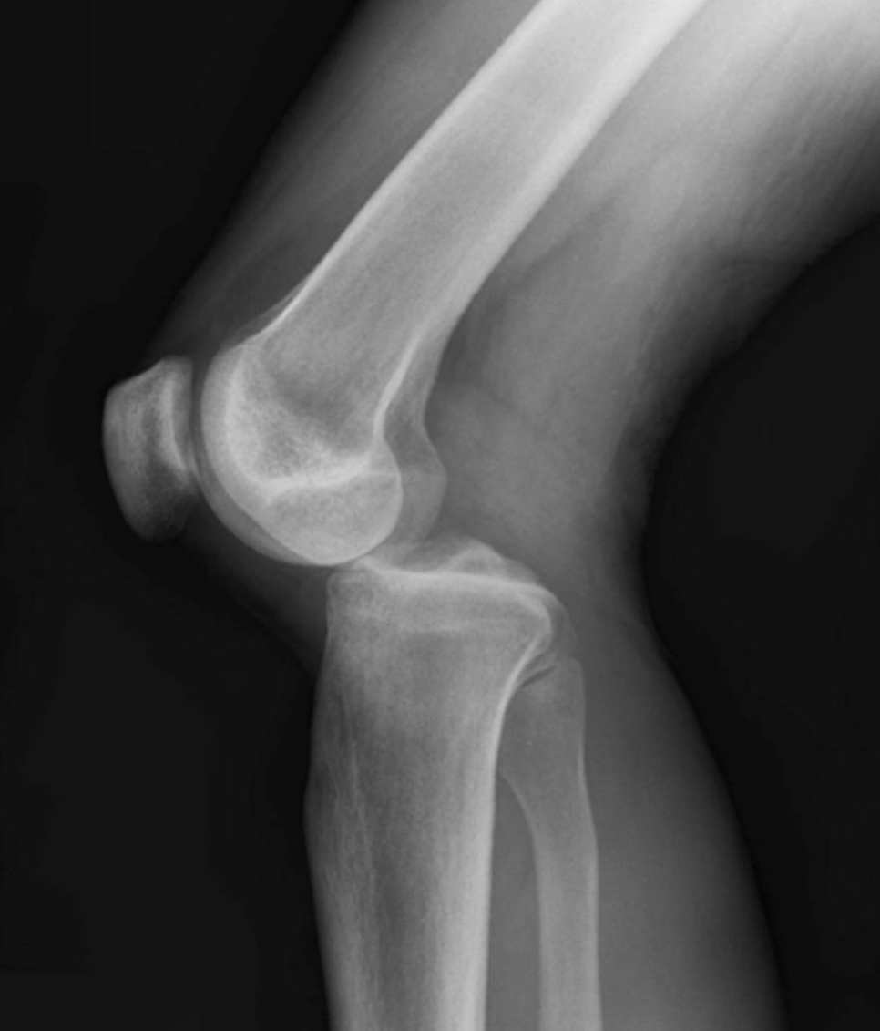
Knee Dislocation Wikipedia from upload.wikimedia.org Magnetic resonance imaging (mri) interpretation of the knee is often a daunting challenge to the student or physician in training. Seems like it should be pretty easy, right? The knee joint is the junction of the thigh and leg. The main knee muscles are the quadriceps, hamstrings and calf muscles. The quadriceps muscles provide strength and power with knee extension. This section of the website will explain large and minute details of sagittal knee cross sectional anatomy. Learn about the muscles, tendons, bones, and ligaments that comprise the knee joint anatomy. Articular surface of patella and femur, condyle, epicondyle and muscles (popliteus anatomy of the ankle and foot in mri:
Related posts of knee muscle anatomy mri muscle anatomy buttocks.
Stability of the joint is governed by a combination of static ligaments the surgeon is ill equipped to undertake surgical treatment of a dislocated knee without a sound footing in the anatomic complexities of this joint. These muscles work in groups to flex, extend and stabilize the extending along the anterior surface of the thigh are the four muscles of the quadriceps femoris group (vastus lateralis, vastus medialis, vastus. Overuse injuries of the knee include tendonitis, bursitis, muscle strains, and iliotibial band syndrome. Involved early gray = muscle: General anatomy and musculoskeletal system. Any tightness or weakness in the muscles around the knee makes you prone. Scroll using the mouse wheel or the arrows. The knee joint is most significantly affected by two major muscle groups: To begin, we use a coronal scan of a left knee. Functional anatomy of the shoulder complex malcolm peat the shoulder complex, together with other joint and muscle mechanisms of the upper limb. This mri knee cross sectional anatomy tool is absolutely free to use. Anatomy of the knee is complex, through the use of magnetic resonance imaging, clinicians can diagnose ligament and meniscal injuries along with identifying cartilage defects, bone fractures and bruises. On anatomical parts the user.


0 Komentar